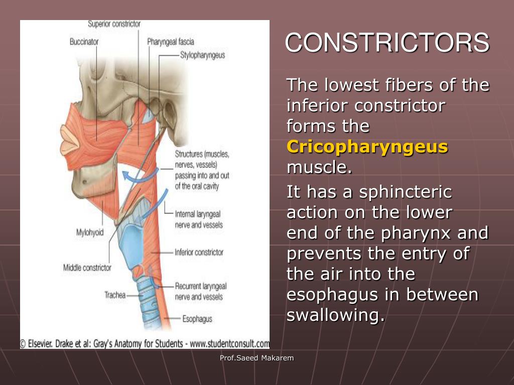

However, the remaining muscles (constrictor and longitudinal groups) emerge from the fourth and sixth arches. The third pharyngeal arch gives rise to the stylopharyngeus muscle. The musculature of the pharynx develops from three pharyngeal arches, i.e., the third, fourth, and sixth arches. The whole pharynx develops from this apparatus, which is present on either side of the developing head. The apparatus consists of arches, pouches, clefts, and membranes which contribute to the development of the head and neck. Embryologyĭuring the fourth and fifth weeks of gestation (development), on either side of the developing pharynx, the formation of the pharyngeal (branchial) apparatus takes place. Air passes from the upper two parts of the pharynx (nasal and oral) and enters the laryngeal pharynx toward the inlet of the larynx and from there to trachea down the respiratory tract. Simultaneously, the epiglottis (single cartilage at the upper part of the larynx) is pushed anteriorly to close the laryngeal inlet preventing food from entering the airways. Lastly, the laryngeal pharynx receives bolus from the oral pharynx and passes it into the esophagus for digestion. These tubes are connected to the middle ears (tympanic cavities) posteriorly and mainly serve to equalize pressure and facilitate the drainage of the secretions of the middle ears. Second, the oral pharynx is a continuation of the oral cavity and functions to pass the bolus toward the laryngeal pharynx below.Īs the bolus passes from the oral cavity, the muscles of the soft palate contract to close the choanae so that food does not enter the nasal cavity. Also, within the lateral surface of the back of the nasopharynx, there are two openings, one on either side, which is called the auditory tubes (Eustachian tubes or pharyngotympanic tubes) surrounded by elevations of mucous membrane called tubal elevations. Regionally, the pharynx divides into three parts which are from superior to inferior:-the nasal pharynx, located behind the posterior nasal apertures (choanae), the oral pharynx, located behind the opening of the oral cavity, and the laryngeal pharynx, located behind the inlet (opening) of the larynx. First, the nasal pharynx is only related to the respiratory tract as air passes through it from the nasal cavities. Knowing its structure and function, embryology, neurovascular supply, musculature, surgical implications, and clinical significance will permit us to comprehend its importance.

Its muscular-membranous integrity allows it to mediate several vital functions related to either organ system, e.g., food swallowing, air conduction, and voice production. It is funnel-shaped with its upper end being wider and located just below the lower surface of the skull, and its lower end is narrower and located at the level of the sixth cervical vertebra (C6) where the commencement of the esophagus posteriorly and the larynx anteriorly takes place. It is the main structure, in addition to the oral cavity, shared by two organ systems, i.e., the gastrointestinal tract (GIT) and the respiratory system. The pharynx is a conductive structure located in the midline of the neck.


 0 kommentar(er)
